01 · PRIMARY
Amino acid sequence
The order of amino acids in a polypeptide.
Sequence, not a particular 3D shape.SEQUENCE → INTERACTIONS → SHAPE → FUNCTION
Explore how a protein folds in water—and what can disrupt its structure.
They describe different aspects of protein structure—not four separate kinds of protein.
01 · PRIMARY
The order of amino acids in a polypeptide.
Sequence, not a particular 3D shape.02 · SECONDARY
α-helices and β-pleated sheets within a chain, stabilised by backbone hydrogen bonds.
Local arrangements of the backbone.03 · TERTIARY
The complete three-dimensional arrangement of one polypeptide, including its local folds.
One whole folded polypeptide.04 · QUATERNARY
Two or more polypeptide subunits associate in one protein assembly.
Each subunit has its own N and C ends.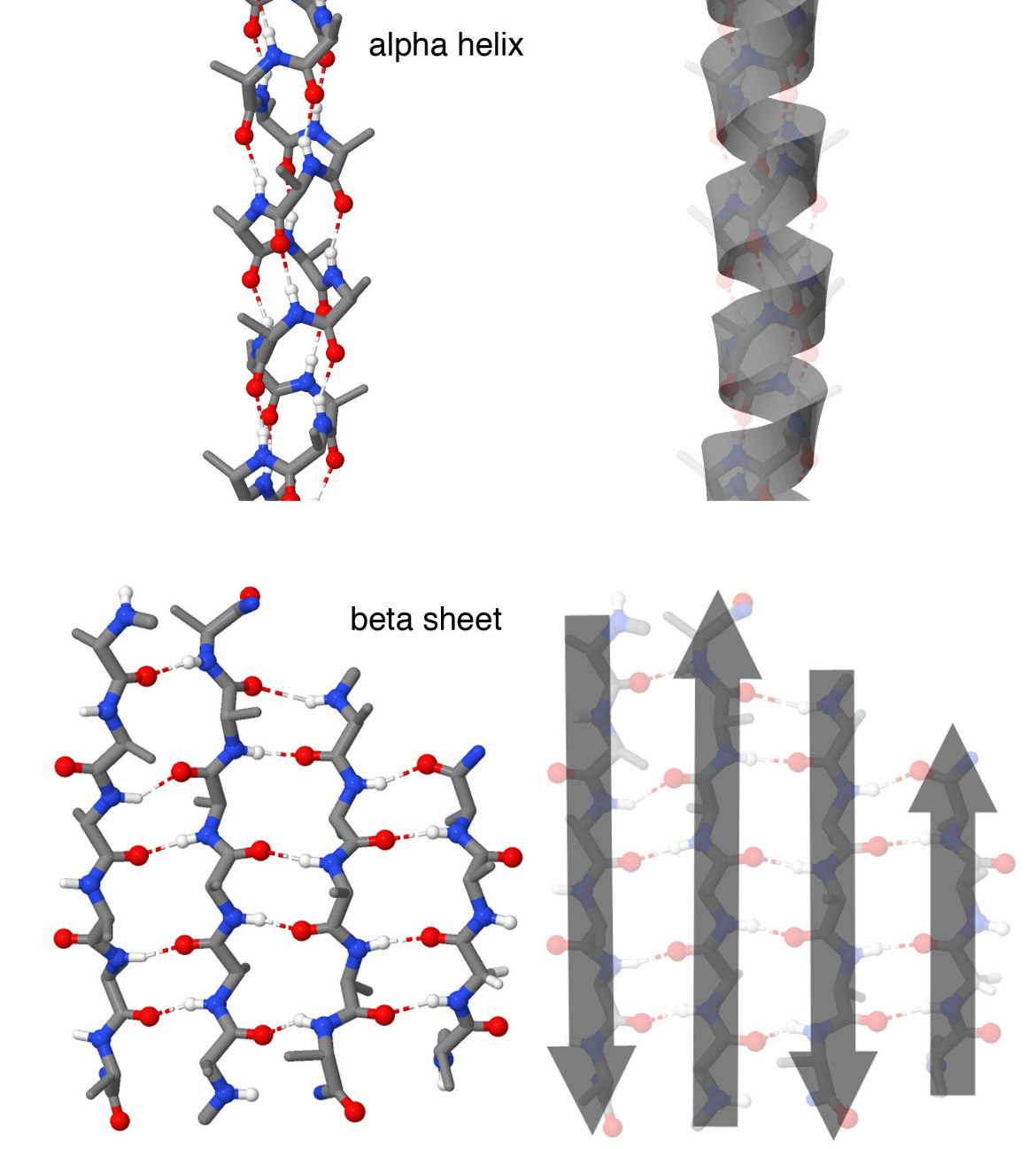
Primary: amino acid sequence, linked by peptide bonds.
Secondary: local α-helices and β-pleated sheets, stabilised by hydrogen bonds between backbone atoms.
Tertiary: the overall shape of one polypeptide, stabilised by hydrophobic interactions, hydrogen bonds, ionic interactions and, in some proteins, disulfide bridges.
Quaternary: interactions among multiple polypeptides. The quaternary investigation uses a separate three-subunit model; each coloured chain has its own N and C ends. Not all proteins have quaternary structure.
The numbered beads represent amino acid residues, and their colours represent R-group properties. The model is a schematic view of a protein in water; it is not an atomic structure or a molecular dynamics simulation.
Helix and sheet views are alternative teaching examples, not a claim that this exact short sequence adopts both structures. Folding paths, contacts and the effect of changing residue 5 are predefined teaching examples, not predictions for an arbitrary sequence. Not every substitution changes folding or function. No real temperature threshold or activity percentage is inferred.
Dashed contacts illustrate selected tertiary interactions. The local-folding investigation shows schematic secondary structures and an enlarged backbone hydrogen bond. Disulfide bridges are not drawn. Hydrophobic interactions are not covalent bonds. Temperature treatment here does not cleave peptide bonds or disulfide bridges. Recovery is shown for this reversible example; some proteins do not recover after denaturation.
CED: 1.7.A.1–6, printed pp. 40–41. Environmental denaturation and possible recovery connect to 3.2.A.1–2, printed p. 69. Detailed amino acid structures and folding calculations are outside the assessed tasks.